DIC is used for imaging live and unstained biological samples, such as a smear from a tissue culture or individual water borne single-celled organisms. Its resolution and clarity in conditions such as this are unrivaled among standard optical microscopy techniques.
What is the principle of interference microscopy?
Interference microscopy uses a prism to split light into two slightly diverging beams that then pass through the specimen. It is thus based on measuring the differences in refractive index upon recombining the two beams. Interference occurs when a light beam is retarded or advanced relative to the other.
What is the principle of phase contrast microscopy?
Principle of Phase contrast Microscopy When light passes through cells, small phase shifts occur, which are invisible to the human eye. In a phase-contrast microscope, these phase shifts are converted into changes in amplitude, which can be observed as differences in image contrast.
What is the principle of and principal use for the phase contrast microscope?
Working principle The basic principle to make phase changes visible in phase-contrast microscopy is to separate the illuminating (background) light from the specimen-scattered light (which makes up the foreground details) and to manipulate these differently.
What are the advantages of phase contrast microscopy and differential interference contrast microscopy?
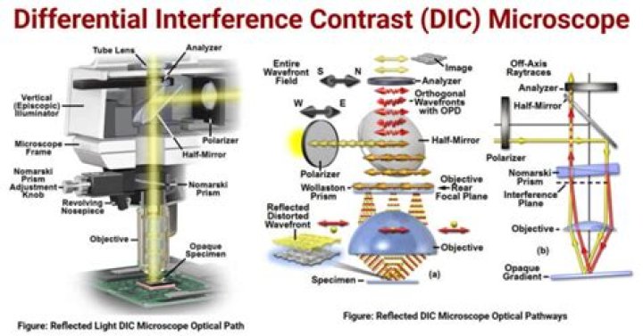
A primary advantage of differential interference contrast over phase contrast is the ability to utilize the instrument at full numerical aperture without the masking effects of phase plates or condenser annuli, which severely restrict the size of condenser and objective apertures.
What are the uses of interference microscope?
Interference microscopy is an optical microscopy technique that uses interference between two white-light illumination beams or rays to generate an image with enhanced contrast.
What is the principle of interference of light?
The interference effect is observed because light reflected from the inner surface of the bubble must travel farther than light reflected from the outer surface, and variations in the soap film thickness produce corresponding differences in the distances light waves must travel to reach our eyes.
What are some possible applications for interference of light?
The phenomenon of light-wave interference with oily or filmy surfaces has the effect of filtering light, and, thus, has a number of applications in areas relating to optics: sunglasses, lenses for binoculars or cameras, and even visors for astronauts.
What are the applications of phase contrast microscope?
Phase contrast is by far the most frequently used method in biological light microscopy. It is an established microscopy technique in cell culture and live cell imaging. When using this inexpensive technique, living cells can be observed in their natural state without previous fixation or labeling.
What are the application of electron microscope?
Applications. Electron microscopes are used to investigate the ultrastructure of a wide range of biological and inorganic specimens including microorganisms, cells, large molecules, biopsy samples, metals, and crystals. Industrially, electron microscopes are often used for quality control and failure analysis.
What is the application of phase contrast microscope in life sciences?
The Phase Contrast Microscope is used to visualize unstained living cells. Most of the stains or staining procedures will kill the cells. Phase contrast microscopy enables the visualization of living cells and life events. The Phase Contrast Microscope was developed by Zernike in early 1930s.
What are some advantages of DIC microscopy?
There are numerous advantages in DIC microscopy as compared to phase contrast microscopy. With DIC, it is possible to make fuller use of the numerical aperture of the system and to provide optical staining (color). DIC also allows the microscope to achieve excellent resolution.
What is differential interference?
Differential interference contrast (DIC) microscopy, also known as Nomarski interference contrast (NIC) or Nomarski microscopy, is an optical microscopy technique used to enhance the contrast in unstained, transparent samples.
What is the importance of contrast in microscopy?
2 Answers. Contrast is everything in microscopy. By definition, microscopy involves viewing objects that can’t be seen by the naked eye- by applying contrast, these objects come into view and can be observed at close quarters. Contrast is required in the three main areas of microscopy which are optical, electron and scanning probe.
What does contrast mean in microscopy?
Wiktionary (0.00 / 0 votes)Rate this definition: phase contrast microscopy(Noun) A form of light microscopy in which small differences in refractive index of a transparent specimen are converted into amplitude (contrast) differences in the final image.
What does microscopy, interference mean?
Interference-microscope meaning An instrument used to study the differences in the phase of light transmitted through a specimen.